

How Long Does It Take For Myelin Sheath To Repair
Oct. 11, 2016
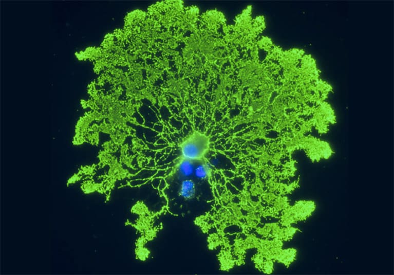
Myelin is essential to the conduction of nerve impulses in the brain and spinal cord, and myelin loss is a key pathophysiological component of neurological injury and disease, including multiple sclerosis, traumatic encephalon and spinal cord injury, stroke, and some neuropsychiatric disorders.
Identifying factors that encourage product of and protect the function of myelin-producing cells — oligodendrocytes and their progenitors (OPCs) — is an important avenue of enquiry aimed at promoting central nervous system (CNS) wellness.
In a recently published article in Biochimica et Biophysica Acta (BBA) Molecular Basis of Disease, Isobel A. Scarisbrick, Ph.D., director of the Neuroregeneration and Neurorehabilitation Laboratory at Mayo Clinic's campus in Rochester, Minnesota, and co-authors investigate the relationship between fat intake, exercise and myelin product in mice.
The roles that dietary fat intake and other external factors play in the production of oligodendrocytes and OPCs is not well-understood. Although brain lipids take high fat content, consumption of a diet containing excess fats and sugars has been shown to exist detrimental to CNS office. Nevertheless, myelin assembly requires a significant amount of lipids, and lipids play an of import role in glial cell myelination.
Practise has been shown to have positive effects on CNS role. Recent inquiry has yielded evidence that exercise can modulate the activeness of diet on the CNS. Additional animal and man studies take shown that new myelin formation in the brain is required to acquire new skills, whether it'south running on an exercise cycle or learning to play the piano or juggle.
"Our study was designed to provide a clearer picture of the interaction between high fatty consumption and exercise training and their effect on myelin and myelin-forming cells in the adult spinal string," explains Dr. Scarisbrick.
Study methods
Mayo researchers studied adult mice, randomized into four experimental groups: Two groups had a sedentary lifestyle and costless access to a regular diet (SRD) or a loftier-fatty diet (SHF). Another ii groups were assigned to either the regular nutrition (ERD) or the high-fat diet (EHF) and had gratis access to an exercise running cycle.
Study findings
After seven weeks, researchers analyzed the lumbosacral spinal cord tissue to measure out the effects of diet and exercise on several building blocks required for myelin assembly in the four report groups, including proteolipid protein (PLP) and myelin basic poly peptide (MBP).
- Loftier-fat diet in combination with exercise grooming increases myelin protein expression. PLP and MBP levels were highest in the group that exercised and consumed a high-fat diet. Exercise training or high fatty consumption lonely also increased PLP. MBP levels in the ERD and the SHF groups were not significantly dissimilar.
- Loftier-fat diet alone or in combination with exercise has the greatest effect on myelin-related protein expression. The SHF and EHF groups had the highest levels of PLP RNA. Elevations in PLP RNA induced by a high-fat nutrition alone (SHF) were significantly greater than those achieved by practice preparation alone (ERD).
- Exercise training protects against loss of OPCs or mature oligodendrocytes induced past a high-fat nutrition. The SHF group had xxx to 50 pct fewer OPCs. While practise solitary didn't bear upon the OPC or oligodendrocyte numbers, mice in the EHF grouping did not experience OPC loss.
- Do training in combination with a high-fat nutrition positively modulates expression of IGF-I levels. This growth gene is known to play important roles in OPC proliferation, survival and differentiation. IGF-one RNA levels were highest in the ERD and EHF groups, suggesting that practice exerts the greatest influence here.
- High-fat diet in combination with exercise converges on energy biosensing systems to restore mitochondrial function and free radical homeostasis. While SHF mice experienced excessive mitochondrial activeness and free radical product, EHF mice upregulated silent mating type information training (SIRT1) and the production of gratis radical scavengers that may protect myelin and myelinating cells from harm in the central nervous system.
These findings suggest that the central nervous organization is capable of adapting to the demands of a high-energy Western diet when afforded aplenty practise.
"Our results suggest that consuming loftier levels of saturated fat in conjunction with a sedentary lifestyle tin lead to a reduction in myelin-forming cells. But exercise grooming can assist reverse this process and promote the myelinogenesis necessary to run into increased energy demands," explains Dr. Scarisbrick.
For more information
Yoon H, et al. Interplay between exercise and dietary fat modulates myelinogenesis in the cardinal nervous system. Biochimica et Biophysica Acta (BBA) Molecular Basis of Illness. 2016;1862:545.

Source: https://www.mayoclinic.org/medical-professionals/physical-medicine-rehabilitation/news/analyzing-the-role-of-diet-and-exercise-in-myelin-production/mac-20429394
Posted by: darlinghicen1943.blogspot.com
0 Response to "How Long Does It Take For Myelin Sheath To Repair"
Post a Comment